
Rupture and Avulsion Tears of the Adductor Longus
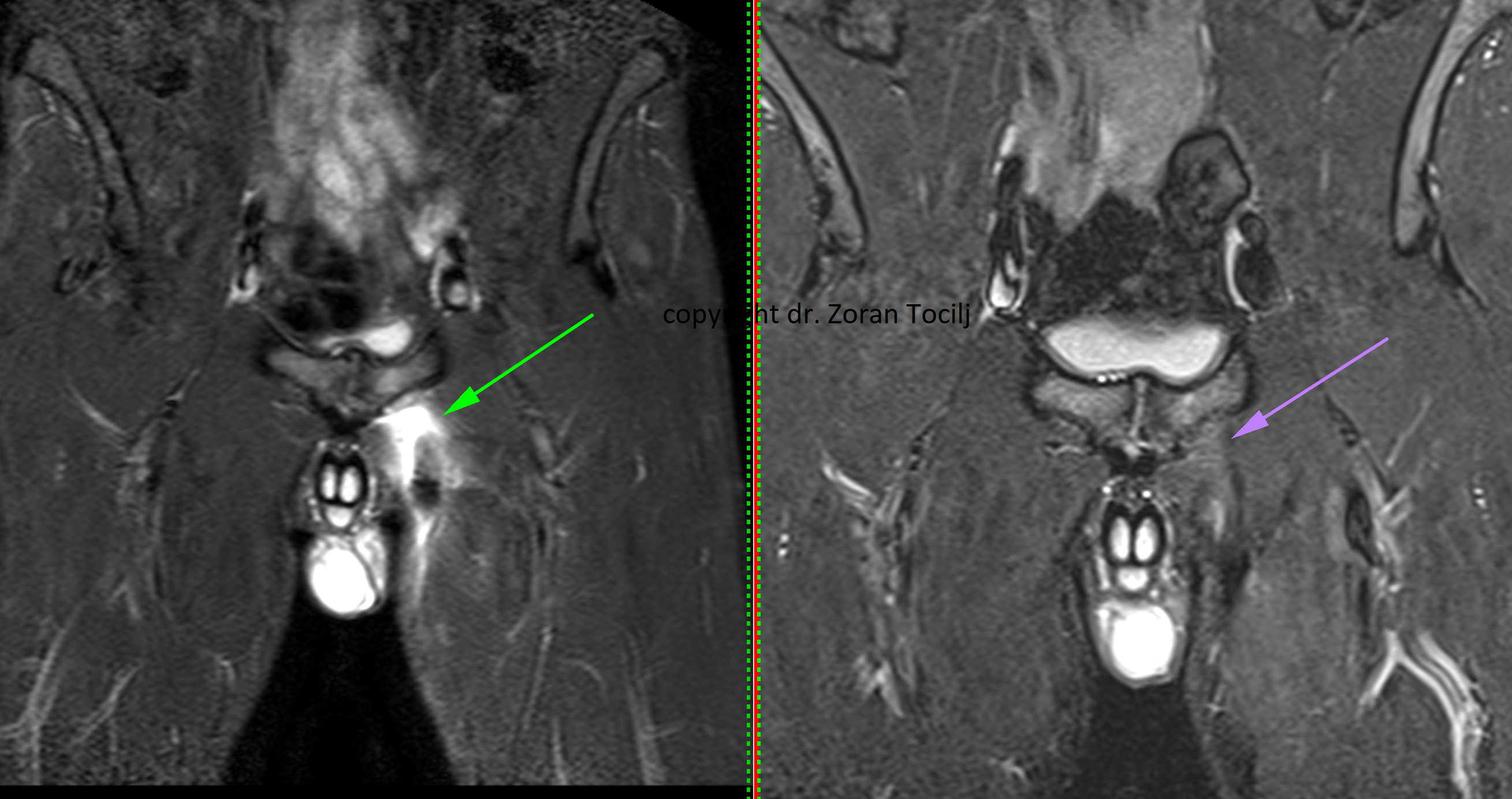
Adductor longus ruptures are relatively common injuries, particularly among football players. According to UEFA statistics, adductor-related injuries account for approximately 9% of all musculoskeletal injuries in professional male football.[1]
In soccer, from a biomechanical perspective, severe adductor longus injuries most commonly occur when a player attempts to reach the ball with the opposite leg. During this movement, the injury occurs in the supporting leg.[2] The adductor longus is subjected to rapid eccentric loading while the muscle is simultaneously activated. Generally, any combination of hip extension, abduction and external rotation produces considerable tensile forces at the proximal tendon and its attachment to the pubic bone, for example during sprinting, shooting and jumping.
In my clinical experience, part of the explanation for the development of an avulsion injury is associated with chronic inflammation of the pubic bone and the surrounding enthesis that has not been adequately treated. The tendon is anchored to the bone through a fibrocartilaginous transition, with specialised collagen fibres commonly referred to as Sharpey’s fibres. In the presence of chronic inflammation, these fibres may gradually deteriorate, lose their structural organisation and become mechanically weaker.
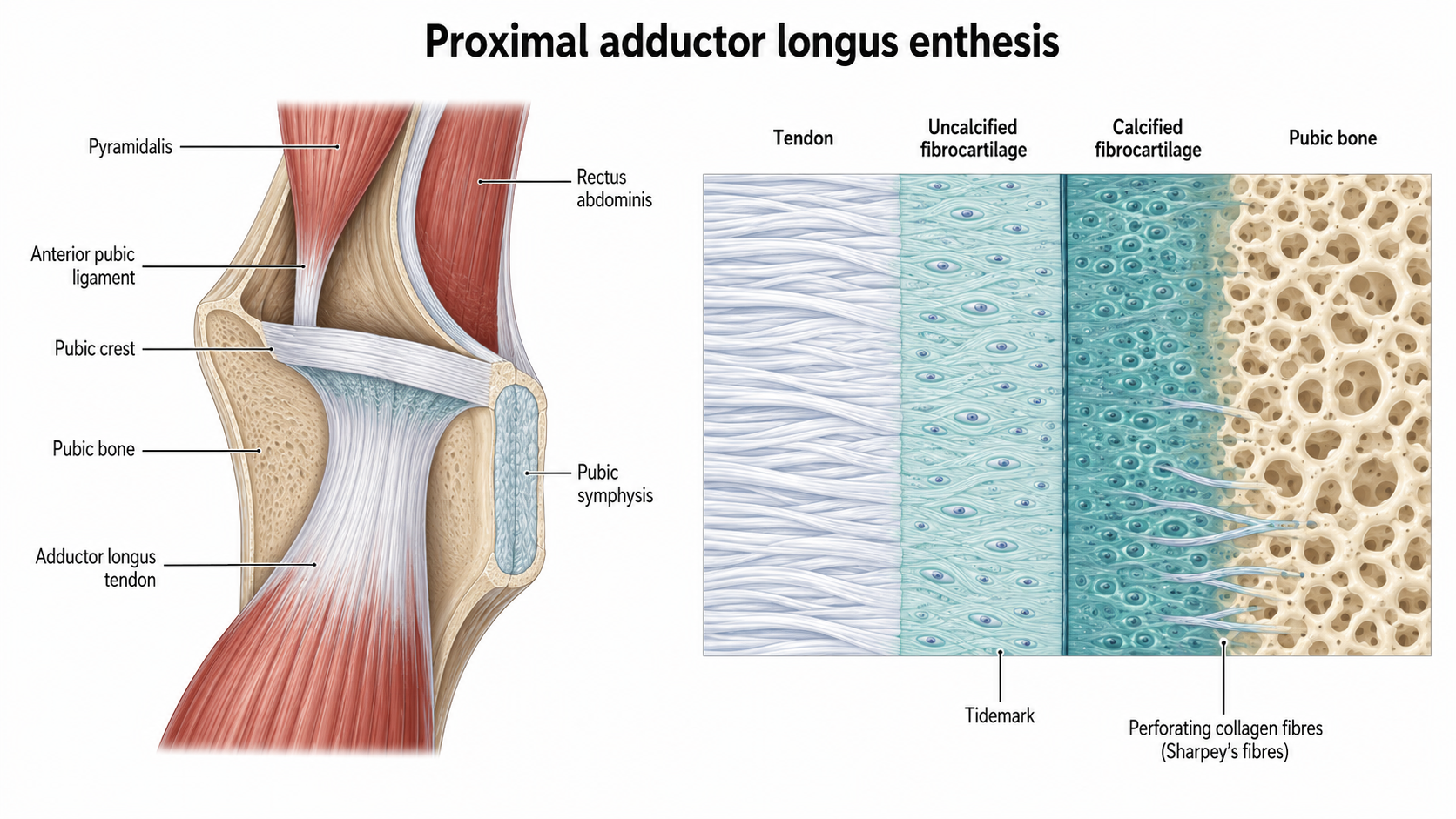
When the already compromised attachment is subsequently exposed to peak traction forces, the remaining collagen fibres fail. Depending on the extent of this failure, the result may be either a partial or a complete rupture of the tendon at its pubic attachment.
Operative Versus Conservative Treatment
Although surgical reattachment may initially appear to be the most logical treatment, it is, in practice, often a slower and less favourable option. A considerable number of published articles and clinical observations support this conclusion.
The recovery period is generally shorter with conservative treatment.[3] Conservative management also avoids the potential adverse effects and complications associated with surgery, including infection, scar formation, adhesions, local irritation, postoperative pain and complications related to the fixation material.
One-year follow-up findings indicate that nonoperatively treated athletes generally report better subjective recovery, good functional outcomes and an absence of persistent pain when compared with surgically treated athletes.[4][5] Conservative management can therefore allow the athlete to regain strength and return to the previous level of sporting activity without the additional tissue trauma caused by surgical intervention.
Therapeutic Approach
Regenerative treatment methods can significantly accelerate tendon recovery, improve the quality of the newly formed tissue and reduce excessive scar formation.
The biological objective of regenerative treatment is not simply to close the defect with fibrotic tissue. The aim is to stimulate the formation, alignment and maturation of new collagen fibres and gradually restore a tendon-like structure capable of transmitting high mechanical loads.
The application of platelet-rich plasma and similar preparations is not always the most appropriate solution. A high local aggregation of platelets may stimulate excessive deposition of disorganised collagen and the formation of fibrotic scar tissue. This scar tissue may provide initial mechanical continuity but may subsequently fail to remodel into a true tendon structure with the normal parallel orientation, elasticity and load-bearing properties of healthy tendon tissue.
Local Prolotherapy
In my therapeutic protocol, locally administered prolotherapy is combined with oral vitamin supplementation, inhalation therapy with oxygen and molecular hydrogen, and a carefully selected combination of physical treatment modalities, including laser therapy, TECAR therapy, electrotherapy, red-light therapy and therapeutic ultrasound.
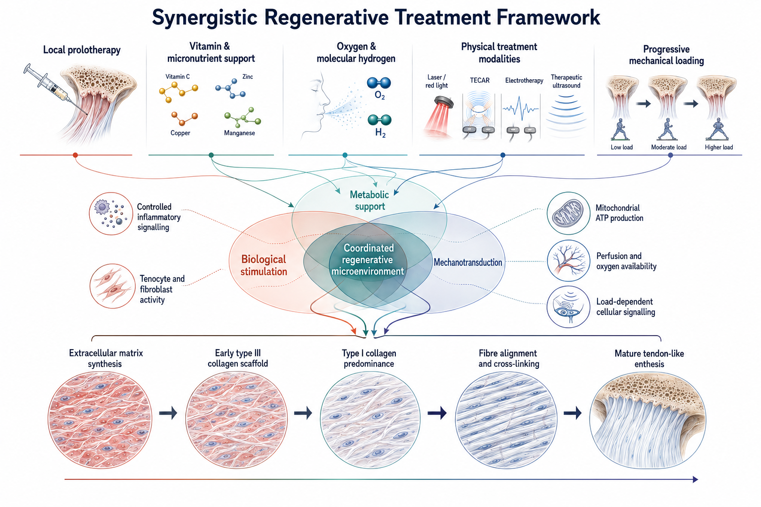
In practical terms: The individual treatment components are designed to work together by combining biological stimulation, metabolic support and progressively controlled mechanical loading.
Local prolotherapy is intended to initiate a controlled regenerative response within the damaged tendon–bone interface. This local stimulation activates tenocytes and supports the production of extracellular matrix components, particularly type I and type III collagen. During the initial phase of healing, type III collagen forms a temporary structural framework. With adequate biological stimulation and progressive mechanical loading, this immature matrix should gradually be remodelled and replaced by stronger, more organised type I collagen.
Vitamin and Micronutrient Support
Adequate oral vitamin and micronutrient supplementation supports the enzymatic processes required for collagen synthesis and maturation. Vitamin C is an essential cofactor for proline and lysine hydroxylation, while zinc, copper, manganese and other micronutrients participate in collagen cross-linking, antioxidant protection and tissue-remodelling processes.
Oxygen Inhalation
Oxygen inhalation is used to improve systemic and local oxygen availability. Oxygen is required for cellular energy production, fibroblast activity, collagen hydroxylation and the development of new microvascular networks within the regenerating tissue.
Molecular Hydrogen
Molecular hydrogen is used for its proposed selective antioxidant and anti-inflammatory effects. Its purpose is to reduce excessive oxidative stress without completely suppressing the controlled inflammatory signalling required for tissue regeneration.
Physical Treatment Modalities
Physical treatment modalities are specially programmed to support prolotherapy and differ from standard everyday treatment programmes. These therapies provide additional biological and mechanical stimulation through increased extracellular blood flow and intracellular mitochondrial activation. Laser and red-light therapy are intended to support mitochondrial activity, ATP production, cellular signalling and local microcirculation. TECAR therapy may improve tissue perfusion and metabolic exchange, while electrotherapy can assist neuromuscular activation and prevent excessive loss of muscle function during periods of reduced loading. Therapeutic ultrasound provides controlled mechanical stimulation that may support fibroblast activity, collagen organisation and local tissue remodelling, while mechanically assisting the body in removing tissue debris.
These treatments are combined with a progressively structured rehabilitation programme. Controlled mechanical loading at a very specific stage of rehabilitation is essential because collagen fibres must be oriented along the direction of functional force transmission. Excessive or premature loading may disrupt newly formed tissue, whereas insufficient loading may result in a weak, disorganised and excessively fibrotic repair.
When the biological and mechanical components of treatment are properly coordinated, the quality of recovery can be significantly improved. In many cases, the tendon may regenerate completely, with restoration of continuity and progressive normalisation of its structure to the extent that the original rupture site is no longer visible on follow-up imaging.
Additional pre- and post-treatment imaging comparisons can be viewed in our stories.
References
- Werner J, Hägglund M, Ekstrand J, Waldén M. Hip and groin time-loss injuries decreased slightly but injury burden remained constant in men’s professional football: the 15-year prospective UEFA Elite Club Injury Study. Br J Sports Med. 2019;53(9):539–546. doi:10.1136/bjsports-2017-097796.
- Jokela A, Pasta G, Della Villa F, et al. Mechanisms of severe adductor longus injuries in professional soccer players: a systematic visual video analysis. Orthop J Sports Med. 2025;13(2):23259671241309647. doi:10.1177/23259671241309647.
- Serner A, Hölmich P, Arnaiz J, Tol JL, Thorborg K, Weir A. One-year clinical and imaging follow-up after exercise-based treatment for acute complete adductor longus tendon avulsions in athletes: a prospective case series. Am J Sports Med. 2021;49(11):3004–3013. doi:10.1177/03635465211015996.
- Farrell SG, Hatem M, Bharam S. Acute adductor muscle injury: a systematic review on diagnostic imaging, treatment, and prevention. Am J Sports Med. 2023;51(13):3591–3603. doi:10.1177/03635465221140923.
- Migliorini F, Maffulli N, Eschweiler J, Tingart M, Baroncini A. Surgical versus conservative management of traumatic proximal adductor longus avulsion injuries: a systematic review. Surgeon. 2022;20(2):123–128. doi:10.1016/j.surge.2021.01.015.