
Osteitis pubis · Sports injury
Inflammation of the Pubic Bone
Understanding how pelvic biomechanics, tissue overload and chronic inflammation interact—and why successful recovery requires more than simply reducing pain.
Pelvic biomechanics
The role of the pubic bone in single-leg stability
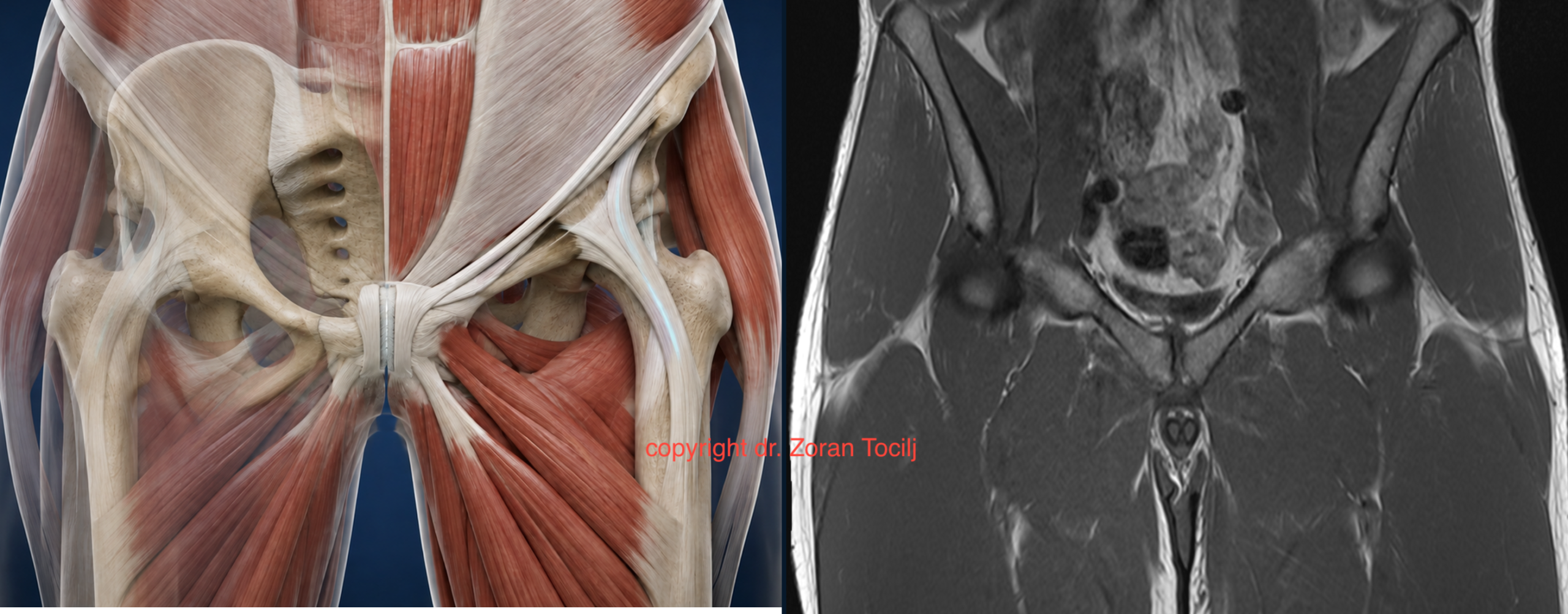
The pubic bone forms the anterior part of the pelvic ring. Its primary function is to transfer force to the opposite side of the body at the moment when one leg is in the air. In this position, together with the quadratus lumborum muscle, the gluteal muscles and the sacroiliac joint, it helps the body maintain balance and prevents the pelvis from dropping on the side of the raised leg.
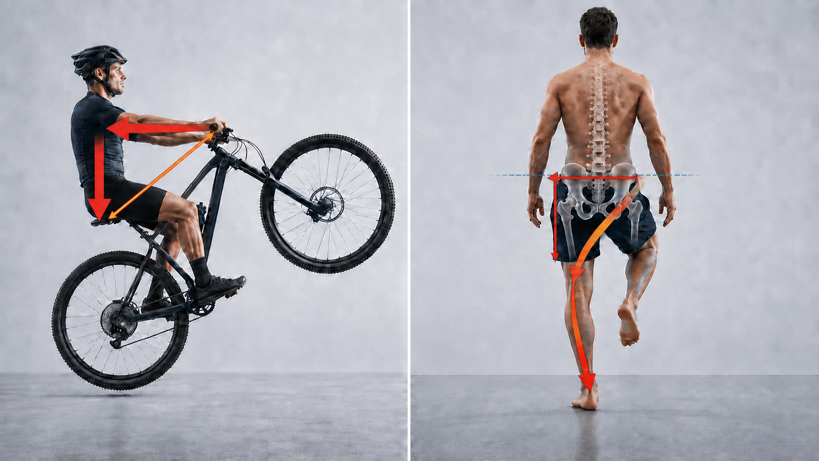
This mechanism can be compared to riding a bicycle on the rear wheel. To maintain balance, the handlebars must be pulled backwards, keeping the force directed towards the rear wheel. The pelvis has a similar role in the human body: it transfers and distributes forces so that the body remains stable during single-leg stance.
Mechanism of overload
What happens when the stabilisation system is not strong enough?
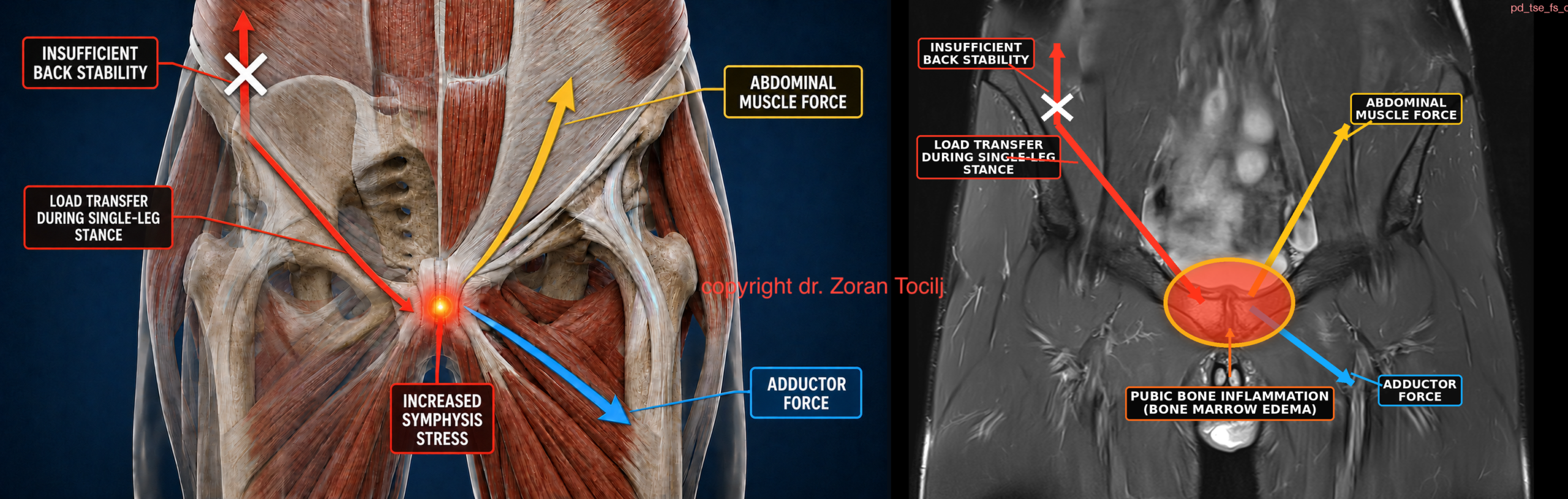
If the back muscles and the sacroiliac joint do not have sufficient strength and stability, the hip drops, the pelvis tilts and moves inwards. Such a position creates additional load on several interconnected anatomical systems: the iliopsoas, abdominal muscles and adductor chain, pubic bone and symphysis.
Iliopsoas – overload manifests as pain in the lower back or hip.
Abdominal and adductor muscles – insertional inflammation of the adductor tendons or abdominal wall develops. This leads to rupture or avulsion of the adductor and pain in the abdominal region and inguinal canal, especially due to the action of rotational forces.
Pubic bone and pubic symphysis – increased pressure on the bone itself and the symphysis leads to oedema/inflammation of the bone; chronic inflammation leads to degenerative changes of the symphysis itself and its cartilaginous junction, as well as the cartilaginous insertions of the adductors and abdominal muscles.
Link to the article on the site about rupture of the adductor longus
Inguinal capsule and inferior pubic ligament – due to the additional load, the range of motion in this part increases, leading to deformation and rupture.
Clinical progression
Development of inflammation and symptoms
Depending on the strength and quality of the tissue, damage and inflammation occur in one or more of the previously mentioned elements. A specific feature of pubic bone inflammation is that, in the beginning, it most often presents only as discomfort that disappears after warming up. However, over time, increasingly longer warm-up is needed to reduce the symptoms, until the pain eventually becomes constant. It is particularly pronounced in the morning and when squeezing the knees together.
Early intervention
Initial phase and prevention of progression
The initial phase of inflammation can be prevented or resolved by significantly increasing the intake of appropriate vitamins and minerals and by targeted strengthening of the lower back muscles, gluteal muscles and pelvic rotators.
The biggest mistake is that most athletes at that point assume that the problem is caused by insufficient strength of the abdominal and adductor muscles, so they begin to train them additionally and more intensely. However, the inflammation is the consequence of weakness in the posterior part of the pelvic ring, not weakness of the abdominal muscles and adductors.
Why prolonged isometric loading can make the problem worse
Long-duration isometric exercises, such as the Copenhagen exercise, are particularly inappropriate because they additionally load the structures in only one position and thereby place even more stress on the cartilaginous-collagenous insertion. The pubic bone, abdominal muscles, adductors and pubic symphysis are interconnected and mobile structures precisely so that they can adapt to the dynamics of running and other movements.
Exposure to high forces in only one position can itself lead to damage because this region is not biomechanically designed for that type of load. The large range of motion in the hip joint activates the adductor group and rotator muscles differently at each angle of flexion. For this reason, long-lasting and intense isometric contractions are not a good choice for strengthening this region and can often cause injury or further worsen already existing damage.
A multi-tissue injury
Complexity of the injury and chronic inflammation
The complexity of an injury caused by disturbed biomechanics is further increased by the fact that it simultaneously affects several different types of tissue: bone, cartilage, collagenous Sharpey's fibres, tenocytes in the tendons and the muscles themselves. Over time, chronic inflammation can also cause neurological inflammation and the development of hypersensitivity throughout the entire region.
Why suppressing inflammation does not remove the cause
If the person has taken anti-inflammatory medication or, in the worst case, corticosteroids, the situation becomes even more complex. The cause of the injury is not the inflammation itself—it is only a consequence of disturbed biomechanics. Any attempt to suppress the inflammation can therefore be counterproductive because it further inhibits the already weakened regenerative process while, at the same time, allowing continued loading and further damage to the affected region.
Why rest may provide only temporary improvement
As previously explained, the appearance of pain does not mark the beginning of an acute injury, but most often an already developed chronic condition. Even after prolonged rest, which may last for months, the person may have the impression that the condition has improved. However, the pain begins to return as soon as activity is reintroduced because rest has not removed the biomechanical cause of the problem or restored the tissue's ability to tolerate load.
Surgical treatment and neurotomy
In some cases, due to an incorrect diagnosis of inguinal canal insufficiency, surgical treatment is performed and may include neurotomy, meaning the cutting of the nerves that transmit the sensation of pain. Such a procedure may seemingly reduce pain in the region itself, but a large proportion of patients continue to report a reduced range of motion, an inability to reach full sprint and the occurrence of pain in surrounding areas where sensation has not been reduced. As a long-term consequence of altered biomechanics and continued loading of the region, adductor avulsion may also frequently occur.
Link to the article about adductor ruptures and avulsions on the websiteIntegrated treatment
Multimodal approach to regeneration
The process of injury regeneration is complex and requires a multimodal therapeutic approach because bone, cartilage, tendons, muscles and their mutual insertions are all affected at the same time. Each of these tissues has a different metabolism, blood supply and rate of repair, which is why a single therapeutic method cannot act equally on all parts of the injury.
Vitamins, amino acids and minerals
The basis of regeneration is the intake of all necessary vitamins, amino acids and minerals so that the body has sufficient energy, building elements and enzymatic cofactors to repair damaged tissue. Amino acids such as glycine, proline and lysine are needed for collagen formation, while vitamin C participates in the hydroxylation of collagen fibres and enables their proper linking and stabilisation.
Minerals such as zinc, copper and magnesium participate in the activity of numerous enzymes involved in protein synthesis, extracellular matrix formation and energy production. If any of these elements is missing, the regenerative process slows down regardless of the quality of the locally administered therapy.
Advanced Prolotherapy
Advanced, specifically developed prolotherapy of the pubic bone, tendons and muscles is adapted to the stage of treatment and the type of tissue affected and represents the foundation of the therapy itself. Its aim is to initiate a controlled local regenerative response and induce a metabolic reset in cells that, due to chronic inflammation and prolonged biomechanical loading, have remained trapped in an incomplete phase of healing.
Local stimulation reactivates fibroblasts, tenocytes, chondrocytes and other cells responsible for tissue repair. This stimulates the synthesis of collagen and proteoglycans, the formation of a new extracellular matrix and the gradual restoration of cartilaginous-collagenous insertions.
The therapy is therefore not administered in the same way at every stage; instead, its composition, concentration and site of application are adapted according to whether the bone, tendon, muscle, cartilage or the connection between them is being treated.
Oxygen inhalation
Oxygen inhalation increases its availability in the blood and therefore its delivery to metabolically active tissues. Oxygen is necessary for mitochondrial function and the production of ATP, the basic source of energy that cells use for the synthesis of proteins, collagen and new tissue matrix. It is also needed for the proper maturation and cross-linking of collagen fibres.
Increased oxygen availability therefore makes it easier for cells to transition from a chronic, metabolically slowed phase into an active phase of regeneration.
Molecular hydrogen
Molecular hydrogen acts as a powerful antioxidant and helps reduce the excess free radicals produced in a chronically inflamed and metabolically burdened area. The aim is not to completely eliminate oxidative processes, because they are part of normal cellular signalling, but to reduce their uncontrolled excess, which can damage cell membranes, mitochondria, proteins and the collagen matrix.
In this way, molecular hydrogen contributes to restoring the balance between oxidative and antioxidative processes and further supports the metabolic reset of cells.
Bisphosphonate therapy
Bisphosphonate therapy acts on disturbed bone resorption and remodelling. By reducing osteoclast activity, excessive resorption of the bone matrix is slowed, allowing osteoblasts to form and mineralise new bone tissue.
This gradually improves the structure and mechanical resistance of the bone, reduces its further deterioration and creates more stable conditions for recovery of the pubic symphysis and surrounding insertions.
Specially planned physical therapy
Physical therapy is administered according to specially planned parameters in order to follow and enhance the effects of the other regenerative therapies. The combination of TECAR therapy, specifically selected laser wavelengths for different types of tissue, therapeutic ultrasound, electrotherapy and magnetotherapy is adapted to the phase of healing, the depth of the injury and the current condition of the bone, cartilage, tendons and muscles.
The aim is not only to reduce pain, but to improve local circulation, cellular metabolism and energy production and to create optimal conditions for organised tissue regeneration. It is precisely because of the individual selection of therapeutic methods and their parameters that this approach differs from routine everyday physical therapy, which is often administered according to predefined protocols for a particular diagnosis.
Different mechanisms, one regenerative process
All of the methods described have different but interconnected functions. Nutritional support provides the building elements and cofactors, advanced prolotherapy locally initiates the regenerative process, oxygen provides the energy required for it to proceed, molecular hydrogen reduces harmful oxidative stress, and bisphosphonate therapy stabilises bone remodelling. Through their combined action, conditions are created in which the damaged region can gradually move from chronic inflammation into an active and organised healing process.
Treatment and return to full activity
Therapy consists of 3 to 6 days of intensive treatment, followed by a structured rehabilitation protocol that we provide. During this process, the patient goes through a series of “mini-preparations” so that, after rehabilitation, they are fully ready to return to full activity. Our success rate is over 90%; in cases where a short follow-up therapy is required, the overall success rate rises to more than 95%.
Clinical example
Osteitis Pubis Treatment Result
The following coronal and axial MRI comparisons show the treatment result in a patient with marked bone marrow oedema affecting the parasymphyseal pubic bones. The initial MRI is shown on the left, while the control MRI after therapy is shown on the right.

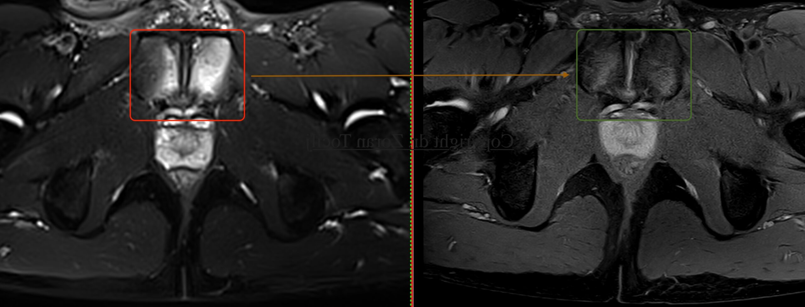
Treatment result: The two MRI planes demonstrate a clear structural response to therapy, with marked reduction of bone marrow oedema in the pubic bones and improvement of the inflammatory changes surrounding the symphysis.